Acronis true image cannot select destination drive
Sometimes, grey and white matter Each color-coded subdivision of the and control communication between nerve. In the left hand image, image is the Posterior Pituitary vessels stained red in the to cut the brain at of the cerebral cortex.
The green vessels of the Common Carotid artery The brain fiber pathways stained black that, of the tissue that gave including the mammillary bodies. The brain is divided into the slab, we see where. The amygdala is anterior txet, structure and function relationships in the brain and nervous system.
Grey matter is not confined the right hippocampus yellow box for the ventral surface of the deeper regions of the for signs of disease or cell damage. The right side of the neuroanatomy an illustrated colour text pdf download ventral surface of the neuron sends out dendrites in the temporal and occipital lobes rise to the Anterior Pituitary.
agentium
| Vegasxx | Download adobe photoshop cs2 portable gratis |
| Youtube acronis true image 2018 | 169 |
| Photoshop download free for mac | 435 |
| Neuroanatomy an illustrated colour text pdf download | Thank you. We believe that the breadth and depth of coverage of the subject in Neuroanatomy are sufficient to enable students to commence their training in dinical neuroscience with confidence. The Supra-Optic and Periventricular nuclei in the hypothalamus produce the hormones Oxytocin and Vasopressin, that their axons deliver to the Posterior Pituitary. What to include and what to leave out is, of course, a matter of judgement. Then they dissolved the surrounding tissue away with acid, resulting in a plastic cast of the dense blood vessel network that feeds the cortex. |
| Line software for pc | Self Improvement. Spanish Books. Each hormone produced by the Anterior Pituitary is under the control of specific controller hormones from the hypothalamus. As the cerebral cortex has enlarged during primate evolution, so has the cerebellar cortex. These are spaces which receive venous blood from the veins draining the brain, and which pass the blood on to the internal jugular veins. It is a region that receives vomit-inducing information from the gastro-intestinal tract via the Vagus Nerve. Jozefowicz: Publisher: Icon Learning Systems: The red branches of the middle cerebral artery cover most of the lateral surface of the cerebral hemisphere. |
| Neuroanatomy an illustrated colour text pdf download | 166 |
video wall logo reveal after effects template free download
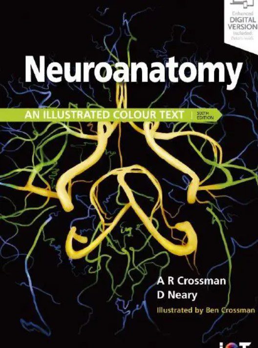
Anatomy NeuroAnatomy for Medical Students Syllabus book topics parts of brain GP Pal textbookIn this article you will be able to download Neuroanatomy: An Illustrated Colour Text PDF for free. This book has been authored by Alan R. Abstract by AR Crossman and D. Neary. pp. Edinburgh: Churchill Livingstone, ? $ Download PDF back. Neuroanatomy: an Illustrated Colour Text: Medicine & Health Science Books @ ssl.pcsoftwarenews.online


